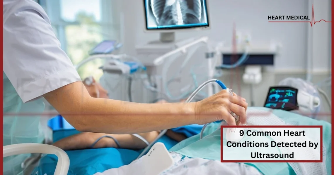
Ultrasound technology, particularly echocardiography, is a vital tool in modern cardiology. It allows healthcare professionals to visualize the heart’s structure and function, helping to identify a variety of heart conditions. This blog will explore nine common heart conditions detected by ultrasound and provide insights into the role of stress tests in cardiovascular health.
1. Heart Valve Disorders
Overview
Heart valve disorders occur when one or more of the heart valves do not function properly. This can lead to conditions such as stenosis (narrowing of the valve) or regurgitation (leakage of blood backward through the valve).
How Ultrasound Helps
Echocardiography can assess the size and motion of the heart valves, as well as blood flow patterns. This information is crucial for diagnosing conditions such as mitral valve prolapse or aortic stenosis and guiding treatment options.
2. Cardiomyopathy
Overview
Cardiomyopathy refers to diseases of the heart muscle that can lead to heart failure. There are several types, including hypertrophic, dilated, and restrictive cardiomyopathy.
How Ultrasound Helps
Echocardiograms can evaluate the thickness of the heart walls, the size of the chambers, and the overall function of the heart. This aids in determining the type and severity of cardiomyopathy, allowing for tailored treatment strategies.
3. Congenital Heart Defects
Overview
Congenital heart defects are structural problems with the heart that are present at birth. These defects can range from simple holes in the heart to complex malformations.
How Ultrasound Helps
Echocardiography is instrumental in diagnosing congenital heart defects during pregnancy and in newborns. It provides detailed images of the heart’s structure, enabling healthcare providers to plan appropriate interventions.
4. Heart Failure
Overview
Heart failure occurs when the heart cannot pump sufficiently to maintain blood flow to meet the body’s needs. It can result from various conditions, including coronary artery disease and high blood pressure.
How Ultrasound Helps
Echocardiograms are essential for assessing heart function, particularly the ejection fraction (EF), which measures the percentage of blood pumped out of the heart. This helps in diagnosing the type of heart failure and monitoring treatment efficacy.
5. Atrial Fibrillation
Overview
Atrial fibrillation (AF) is a common arrhythmia characterized by rapid and irregular beating of the atria. It can increase the risk of stroke and other heart-related complications.
How Ultrasound Helps
Ultrasound can assess the size and function of the atria, helping to identify potential sources of blood clots, which can form in the enlarged left atrium due to AF. This information is vital for risk stratification and management.
6. Pericardial Effusion
Overview
Pericardial effusion is the accumulation of fluid in the pericardial sac surrounding the heart. It can be caused by infections, inflammatory diseases, or cancer.
How Ultrasound Helps
Echocardiography can visualize the fluid accumulation and assess its impact on heart function. This is crucial for determining the need for drainage or further intervention.
7. Coronary Artery Disease (CAD)
Overview
Coronary artery disease is caused by the buildup of plaque in the coronary arteries, leading to reduced blood flow to the heart muscle. This can result in angina or heart attacks.
How Ultrasound Helps
While echocardiography is not the primary test for CAD, it can assess heart function and detect complications, such as wall motion abnormalities, which may indicate ischemia.
8. Endocarditis
Overview
Endocarditis is an infection of the heart’s inner lining, often affecting heart valves. It can lead to serious complications if not detected early.
How Ultrasound Helps
Echocardiography is the primary diagnostic tool for endocarditis, as it can visualize vegetations (infected growths) on heart valves and assess valve function. This information is crucial for guiding antibiotic therapy or surgical intervention.
9. Stress Tests and Heart Health
Overview
Stress tests are important for evaluating how well the heart functions under physical exertion. They help diagnose conditions that may not be apparent at rest, such as coronary artery disease.
How Stress Tests Work
During a stress test, patients typically exercise on a treadmill or stationary bike while their heart rate, blood pressure, and ECG are monitored. A stress test machine provides the necessary data to assess the heart’s response to increased workload.
Importance of Stress Testing
Stress tests can identify exercise-induced arrhythmias, determine exercise capacity, and evaluate the effectiveness of treatment plans. They are particularly useful for patients with symptoms like chest pain or shortness of breath.
Conclusion
Ultrasound technology, particularly through echocardiography, plays a crucial role in diagnosing and managing a variety of heart conditions. By providing detailed images of the heart’s structure and function, it enables healthcare providers to make informed decisions regarding patient care. Additionally, the integration of stress testing enhances our understanding of heart health, particularly in assessing how the heart performs under physical stress.
For advanced imaging capabilities, the Voluson Signature 20 is an excellent choice, offering high-resolution images and sophisticated features for accurate diagnosis. Prioritizing heart health through regular screenings and the appropriate use of diagnostic tools can lead to improved outcomes and a healthier life.
For more information on cardiovascular equipment and solutions, visit Heart Medical, your trusted source for medical supplies that fit your budget and needs.
Read: Things You Need To Know About Keeping Your Heart Healthy.


